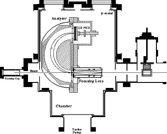
The experimental apparatus, shown in detail in Fig. 1,
is presently in use at the J.R. Macdonald laboratory.
The spectrograph consists
of a large hemispherical analyser with an inner radius ![]() mm and
outer radius
mm and
outer radius ![]() mm, a 40mm diameter 2-dimensional
position sensitive detector (2D-PSD) made up of two MCP's, a
resistive anode encoder (RAE) and a cylindrical 4-element
lens mounted at the entrance of the analyser. The lens
provides a virtual slit by focusing the incoming electrons down to a diameter of
about 1 mm for
improved energy resolution. The actual entrance slit (see Fig. 1)
has a diameter of 6 mm to
allow for the uninhibited passage of the primary ion beam.
The lens is also used for decelerating the incoming
electrons in the high resolution mode of operation.
The hemispherical analyser has an overall acceptance energy range of 20
mm, a 40mm diameter 2-dimensional
position sensitive detector (2D-PSD) made up of two MCP's, a
resistive anode encoder (RAE) and a cylindrical 4-element
lens mounted at the entrance of the analyser. The lens
provides a virtual slit by focusing the incoming electrons down to a diameter of
about 1 mm for
improved energy resolution. The actual entrance slit (see Fig. 1)
has a diameter of 6 mm to
allow for the uninhibited passage of the primary ion beam.
The lens is also used for decelerating the incoming
electrons in the high resolution mode of operation.
The hemispherical analyser has an overall acceptance energy range of 20 ![]() and a mean energy resolution of 1
and a mean energy resolution of 1 ![]() (when the lens is focused under low resolution mode).
Novel
features include the use of an off-center entrance aperture placed at a
non-zero entrance potential
(when the lens is focused under low resolution mode).
Novel
features include the use of an off-center entrance aperture placed at a
non-zero entrance potential ![]() . This has been found to
improve the focusing properties of the spectrometer
by compensating for fringing field effects, while enhancing
the acceptance energy range[2].
. This has been found to
improve the focusing properties of the spectrometer
by compensating for fringing field effects, while enhancing
the acceptance energy range[2].

Figure: The Experimental Setup
Details of the spectrograph features, its operation to date, the electronics in use and the data acquisition system have been briefly reported in [2]. A full theoretical treatment of the electron trajectories in this non-typical hemispherical analyser will be reported elsewhere[4]. As we have not yet finished with the full characterisation of our spectrograph, we do not scan the voltages but keep them fixed on all elements including the lens. In particular, the lens voltages are determined to first order empirically in non-deceleration mode and then held fixed for all electrons with similar initial energies even when decelating. While this does not allow for the best results, it is a first step in exploring the many variable space of this 4-element lens.
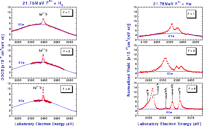
Results are presented in Fig. 2 for two different ion charges which give
rise to different projectile KLL Auger spectra.
The data were corrected for
dead time and were fitted with a Gaussian function superimposed on
a theoretical curve based on the elastic scattering model (Binary Encounter electron (BEe)
peak).
The energy calibration was performed using an electron gun,
while the beam energy was determined with high
accuracy from the known energy of the F ![]() (2p
(2p ![]() line.
The energy resolution is defined as
line.
The energy resolution is defined as ![]() ,
where
,
where ![]() is the initial energy,
is the initial energy, ![]() the pass energy and
the pass energy and ![]() the
deceleration factor. The spectra are very similar in quality with those already obtained
for the same F
the
deceleration factor. The spectra are very similar in quality with those already obtained
for the same F ![]() [5] and F
[5] and F ![]() [6, 7]
collision systems with a conventional tandem spectrometer.
However, our new spectrometer is presently about a factor of 15-20
times more efficient even when having to run with ion beam currents of
about 5nA imposed by deadtime limitations[3]. Typically, beam currents
in zero-degree measurements using conventional tandem spectrometers can be
about 20-100 times larger at 100-500nA depending on the collision system studied.
[6, 7]
collision systems with a conventional tandem spectrometer.
However, our new spectrometer is presently about a factor of 15-20
times more efficient even when having to run with ion beam currents of
about 5nA imposed by deadtime limitations[3]. Typically, beam currents
in zero-degree measurements using conventional tandem spectrometers can be
about 20-100 times larger at 100-500nA depending on the collision system studied.

Figure: High resolution projectile Auger spectra plotted as a
function of the deceleration factor F: [Left] Double differential
cross sections (DDCS) of the ![]() Auger line produced by
RTE in collisions of
21.75MeV F
Auger line produced by
RTE in collisions of
21.75MeV F ![]() H
H ![]() ,
[Right] Normalized yield showing various KLL Auger lines
produced in collisions of
21.78 MeV F
,
[Right] Normalized yield showing various KLL Auger lines
produced in collisions of
21.78 MeV F ![]() + He.
+ He.
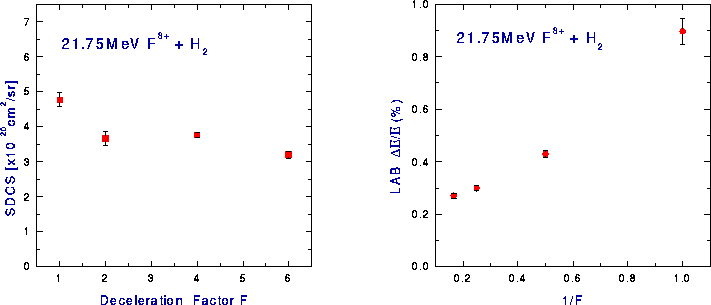
In Fig. 3 we plot the area under the F ![]() (2p
(2p ![]() ) line
(single differential cross section or SDCS) as a function of the deceleration factor.
This should remain constant under correct operation and constitutes one of the basic
tests of any spectrometer. The area is seen to
remain invariant (within the statistical error) for deceleration
factors in the region of 2 up to 6 even in the present way of running the
spectrograph and lens. The average value of the measured SDCS after
transforming it to the projectile rest frame is
) line
(single differential cross section or SDCS) as a function of the deceleration factor.
This should remain constant under correct operation and constitutes one of the basic
tests of any spectrometer. The area is seen to
remain invariant (within the statistical error) for deceleration
factors in the region of 2 up to 6 even in the present way of running the
spectrograph and lens. The average value of the measured SDCS after
transforming it to the projectile rest frame is
![]() cm
cm ![]() /sr
in good agreement with the best measurement to date using the KSU
tandem spectrometer, having a value
of
/sr
in good agreement with the best measurement to date using the KSU
tandem spectrometer, having a value
of ![]()
![]() , and with theory which predicts
a value of
, and with theory which predicts
a value of ![]()
![]() [5].
[5].
In Fig. 3 it is seen that for large deceleration factors the resolution is not anymore proportional to 1/F (i.e. the pass energy). This is probably due to the fact that the lens voltages were kept constant at the value that determined the best focusing (or resolution) conditions for the case of no deceleration (F=1). At larger deceleration factors the size of the image at the entrance of the analyser (exit of the lens) as well as the angle of incidence of the electrons entering the analyser increases with increasing deceleration factor with a corresponding degradation of the energy resolution. Clearly we need to vary the voltages on the lens as the deceleration factor is changed. This will require the full characterization of the lens and will be done in the near future.

Figure: Measurements of laboratory
single differential cross sections (SDCS)
of the ![]() RTE line produced in collisions of
21.75 MeV F
RTE line produced in collisions of
21.75 MeV F ![]() + H
+ H ![]() as a function of deceleration
factor F:
[Left] Variation of the area under the peak (SDCS) with F,
[Right] Variation of relative energy resolution with F.
as a function of deceleration
factor F:
[Left] Variation of the area under the peak (SDCS) with F,
[Right] Variation of relative energy resolution with F.
In conclusion we have shown that it is possible to use a single stage electron spectrometer with a position sensitive detector to perform high resolution measurements in the beam direction, thus minimizing kinematic broadening effects. This is made possible by using a large entrance aperture on a hemispherical analyser which allows for the uninhibited passage of the ion beam, thus avoiding large backgrounds from slit scattering. However, since a small entrance aperture is required for good resolution, a virtual slit was provided by focusing and decelerating electrons before entering the analyser by means of a 4-element lens. The feasibility of this idea is supported by the first test results presented here. Most important, our new apparatus has a much higher gain of about 15-20 over previously used tandem parallel-plate spectrometers and when equipped with a faster data detection/acquisition system a further gain of about 100 should be possible. In the future, when we have finished with the full characterization of our lens, we shall also be able to perform measurements of higher resolution, close to the limit of kinematic broadening.